Microscopy
|
Magnifying Power
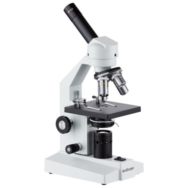
A compound microscope has two sets of lenses. The lens you look through is called the ocular. The lens near the specimen being examined is called the objective. The objective lens is one of three or four lenses located on a rotating turret above the stage, and that vary in magnifying power. The lowest power is called the low power objective (LP), and the highest power is the high power objective (HP). You can determine the magnifying power of the combination of the two lenses by multiplying the magnifying power of the ocular by the magnifying power of the objective that you are using. For example, if the magnifying power of the ocular is 10 (written 10X) and the magnifying power of an objective is 4 (4X), the magnifying power of that lens combination is 40X. |
Field of View (FOV)
The field of view is the maximum area visible through the lenses of a microscope, and it is represented by a diameter. To determine the diameter of your field of view, place a transparent metric ruler under the low power (LP) objective of a microscope. Focus the microscope on the scale of the ruler, and measure the diameter of the field of vision in millimeters. Record this number.
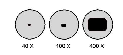
When you are viewing an object under high power, it is sometimes not possible to determine the field of view directly. The higher the power of magnification, the smaller the field of view.
The diameter of the field of view under high power can be calculated using the following equation:
For example, if you determine that your field of view is 2.5 mm in diameter using a 10X ocular and 4X objective, you will be able to determine what the field of view will be with the high power objective by using the above formula. For this example, we will designate the high power objective as 40X.
Estimating the Size of the Specimen Under Observation
Objects observed with microscopes are often too small to be measured conveniently in millimeters. Because you are using a scale in millimeters, it is necessary to convert your measurement to micrometers. Remember that 1 μm = 0.001 mm.
To estimate the size of an object seen with a microscope, first estimate what fraction of the diameter of the field of vision that the object occupies. Then multiply the diameter you calculated in micrometers by that fraction. For example, if the field of vision’s diameter is 400 μm and the object’s estimated length is about one-tenth of that diameter, multiply the diameter by one-tenth to find the object’s length.


